

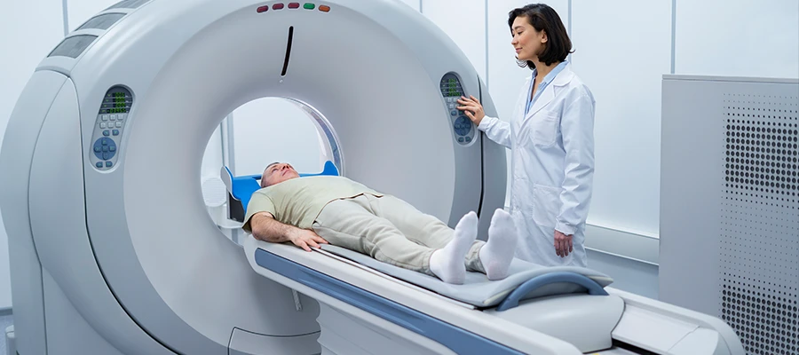
What is an MRI?
MRI, a method that allows for detailed examination of different organs and tissues in the body, is used for diagnostic purposes in modern medicine. Thanks to MRI, doctors can clearly observe structural changes in the brain, liver, kidneys, and joints. This imaging method uses magnetic fields and radio waves and does not involve radiation. The patient usually does not feel pain during the procedure and must remain still. MRI plays a critical role in tumor detection, vascular occlusion, infections, and the evaluation of musculoskeletal problems. Contrast-enhanced MRI provides detailed visualization of vessels and lesions. Non-contrast MRI provides sufficient images in some cases. The procedure usually takes between 20 and 60 minutes, and the patient should remove any metal objects. MRI results provide an important reference in diagnosis and treatment planning and are an indispensable imaging method in modern medicine.
For what purposes is magnetic resonance used?
Magnetic resonance imaging (MRI) is preferred in various medical conditions for the detailed examination of organs and tissues in the body. This method is beneficial in evaluating conditions such as headaches, back pain, post-traumatic tissue damage, and joint complaints. Thanks to MRI, tumors, vascular occlusions, inflammations, and muscle problems can be examined in detail. No radiation is used during the procedure, and it is painless for most patients. Contrast-enhanced MRI allows for clear visualization of lesions and vascular structures. Non-contrast MRI provides sufficient images in some cases. Patients remain motionless inside the device, maintaining their position throughout the scan. MRI offers the possibility of early intervention in the diagnosis and follow-up process and plays a critical role in modern medicine.
How does an MRI machine create images of the body?
The device aligns protons in the body via a magnetic field and records the signals returning as radio waves. These signals are processed by a computer to create detailed organ images. Thanks to MRI, structural abnormalities in areas such as the brain, spinal cord, liver, kidneys, and joints can be clearly examined. Contrast-enhanced MRI makes the details of vessels and lesions more distinct. Non-contrast MRI provides sufficient images in some cases. The patient must remain still during the procedure. MRI imaging is a critical reference for doctors in diagnosis and treatment planning and is safely applied in modern medicine.
For what symptoms do doctors order an MRI?
Headaches, back pain, joint problems, and abdominal pain are among the most common complaints for MRI scans. MRI is also preferred for injuries resulting from trauma and some neurological complaints. This method allows for detailed examination of tumors, vascular occlusions, muscle and joint problems, and inflammations. Contrast-enhanced MRI enables clear observation of lesions and vascular details. Non-contrast MRI provides sufficient images in some cases. The scan time generally ranges from 20 to 60 minutes. The patient remains motionless inside the machine throughout the imaging process.
What health problems can be detected with MRI?
Tumors, vascular blockages, infections, and muscle and joint problems can be detected in detail thanks to MRI. Structural changes in organs such as the brain, spinal cord, liver, and kidneys can be clearly examined with this method. Contrast MRI allows for better visualization of vascular and lesion details. Non-contrast MRI provides sufficient images in some cases. The patient must remain still during the procedure and remove any metal objects. MRI results constitute an important reference for doctors in diagnosis and treatment planning. This method is used safely in modern medicine and provides the opportunity for early diagnosis.
Types of MRI Scans and Their Applications
MRI scan types include contrast-enhanced, non-contrast, functional, diffusion, and perfusion MRI. Contrast-enhanced MRI allows for detailed examination of vascular structure and lesions. Non-contrast MRI provides sufficient images in some cases. Functional MRI is preferred for evaluating organ function. Diffusion MRI is useful in detecting cellular changes. Perfusion MRI is used to monitor blood flow and tissue nutrition. Each type enables the early detection of different health problems. MRI scan types play a critical role in the diagnosis and monitoring process in modern medicine and are widely preferred by doctors.
What does a contrast-enhanced MRI scan mean?
A contrast-enhanced MRI is an imaging technique that uses a special dye to clearly visualize lesions and blood vessels. The contrast agent is administered intravenously, allowing for detailed observation of different tissues within organs. Contrast-enhanced MRI is preferred for conditions such as tumor detection, vascular occlusion, and inflammation. Non-contrast MRI may provide sufficient images in some cases. The patient must remain still during the procedure. Contrast-enhanced MRI results are critically important for diagnosis and treatment planning and are reliably used in modern medicine.
In what situations is non-contrast MRI preferred?
In some cases, contrast medium is not needed, and non-contrast MRI provides sufficient images. Non-contrast MRI is preferred in cases where there is no suspicion of a tumor and vascular detail is not critical. This method is useful in evaluating headaches, back pain, and joint complaints. During the procedure, the patient must remain still and remove all metal objects. Non-contrast MRI does not involve radiation and is painless for most patients. The scan time generally ranges from 20 to 60 minutes, and the results provide an important reference for doctors.
What are the stages of an MRI scan?
Before an MRI scan, the patient must remove all metal objects and share their medical history with the doctor. The patient is placed inside the machine and remains in a still position. The device records the signals returning from protons via magnetic fields and radio waves. The procedure is performed according to whether it is a contrast-enhanced or non-contrast MRI. The signals are transmitted to a computer, and detailed images of organs and tissues are obtained. The procedure usually takes between 20 and 60 minutes. MRI results play a critical role in diagnosis and treatment planning and are used reliably in modern medicine.
Preparations to be Made Before an MRI
It is important for patients to remove all metal objects and to discuss pregnancy and kidney problems with their doctor before an MRI. Comfortable clothing should also be preferred before the procedure. In some cases, it may be necessary to come on an empty stomach because contrast medium will be administered. The patient must remain still inside the machine and maintain their position. No radiation is used during an MRI, and it is painless for most patients. These preparations ensure accurate and clear images during the scan. MRI results provide a critical reference in diagnosis and treatment planning.
How to Choose Between Open and Closed MRI?
MRI machines have different designs, and the choice is made according to the patient’s comfort and imaging needs. Open MRI offers a more comfortable experience for patients with claustrophobia or those who are overweight. Closed MRI is preferred in cases requiring higher resolution and detail. These machines record signals returning from protons in the body using magnetic fields and radio waves. The choice between contrast-enhanced or non-contrast MRI affects image quality depending on the type of scan. The doctor guides the patient’s choice of machine based on their complaints and imaging goals. The patient must remain still during the procedure, and any metal objects must be removed. Open MRI is usually completed quickly, while closed MRI may take longer in some cases. This choice is critical for accurate diagnosis and treatment planning.
What precautions should be taken during an MRI scan?
During an MRI scan, patients should remain still to ensure clear and accurate images. Removing metal objects prevents potential interference with the device’s magnetic field. Conditions such as pregnancy or kidney problems should be reported to the doctor beforehand. If a contrast-enhanced MRI is to be performed, fasting may be necessary. It is important to wear comfortable clothing and inform the doctor about any medications taken before the procedure. The noise produced by the device during the MRI is normal, and earplugs can be used. The procedure is painless and safe for patients. Immobility, correct positioning, and precautions ensure the preservation of image quality and contribute to the reliability of the results for diagnosis.
MRI Prices 2026
In 2026, MRI prices typically range between €1200 and €5000.
Can I eat before an MRI scan?
Light meals generally don’t cause problems before an MRI, but it’s recommended to come on an empty stomach if a contrast-enhanced MRI is to be performed. The doctor will give instructions based on the patient’s health condition and the type of scan. Heavy and fatty meals can increase the risk of stomach upset or nausea. Coming on an empty stomach ensures effective use of the contrast agent and clear images. The doctor should be informed about any regular medication use. The patient should remain still and maintain their position inside the machine during the procedure. Dietary recommendations before an MRI may vary depending on the type of scan and the organ being examined.
Why does noise occur during an MRI?
The coils and gradient systems of the MRI machine vibrate, and this vibration, combined with radio waves, creates noise. This noise is normal and is caused by the operation of the device. Using earplugs or headphones can reduce discomfort. The noise does not affect image quality and does not pose any health risks during the procedure. With immobility and maintaining position, the scan is completed safely. MRI is radiation-free and painless for most patients.
How long does it take to get MRI results?
MRI results are usually evaluated by a radiologist within 1–3 business days of the scan being completed. In urgent cases, some results may be delivered the same day or within a few hours. The images are processed on a computer and interpreted by the doctor. Contrast-enhanced MRIs may slightly prolong the process as they require additional evaluation. The results provide a critical reference for diagnosis and treatment planning. Patients are informed by the clinic about the report delivery time.
Does MRI involve radiation?
MRI scans do not involve radiation and are therefore considered a safe method. The procedure uses magnetic fields and radio waves to record signals reflected from protons. This allows for detailed examination of the brain, kidneys, liver, and joints. Both contrast and non-contrast options do not increase radiation risk. With immobility and maintaining the correct position, the procedure is completed safely. MRI is a widely used diagnostic method in modern medicine.
Why are metal objects removed during an MRI scan?
Metal objects can interact with the strong magnetic field of the MRI machine, posing a safety risk and causing image distortions called artifacts. Therefore, metal objects such as necklaces, watches, earrings, and piercings should be removed before the procedure. Some prostheses or implants are scanned after checking their MRI compatibility. Removing metal objects is critical for safety and obtaining clear images. Immobility and maintaining position, along with image quality, are essential.
Can an MRI be performed on the entire body?
Whole-body MRI allows for the examination of organs and tissues at once and is preferred in specific situations. This method can be used for suspected metastases, systemic diseases, or general screening purposes. Contrast or non-contrast options are determined according to the purpose of the scan. The patient must remain still and remove all metal objects during the procedure. The duration can vary between 45 and 90 minutes, and all organs are examined in detail. Whole-body MRI is safely applied in modern medicine and is a critical method in the diagnostic process.
What does an MRI report mean?
An MRI report is prepared by a radiologist who interprets the images obtained during the scan. The report includes structural changes in organs and tissues, tumors, vascular occlusions, or inflammations. The doctor evaluates the report to make a diagnosis and create a treatment plan. A contrast-enhanced MRI report shows vascular and lesion details more clearly. A non-contrast MRI report provides sufficient information in some cases. The MRI report serves as a critical reference for patients to understand their health status and plan their follow-up process.