

What is Perfusion MRI?
A specialized method has been developed to examine blood flow and tissue nutrition in organs and tissues throughout the body in detail. This method, perfusion MRI, allows for clear observation of the details of vessels and tissues. While contrast-enhanced images reveal details of blood flow and lesions, in some cases, non-contrast perfusion MRI can also provide sufficient information. The patient must remain still during the procedure and remove all metal objects. Perfusion MRI is safely used in the evaluation of diseases such as tumors, vascular occlusions, and strokes. During the scan, the device’s magnetic field and radio waves record signals from protons. By processing the images on a computer, the doctor can perform a detailed analysis. The procedure usually takes 20–60 minutes and is radiation-free. In modern medicine, perfusion MRI plays a critical role in accurate diagnosis and treatment planning. This method allows for a detailed assessment of the patients’ vascular and tissue conditions.
What is perfusion MRI used for?
Perfusion MRI is a preferred imaging method for detailed examination of blood flow and tissue nutrition. Blood circulation and oxygenation in the brain, heart, kidneys, and other organs are clearly observed with perfusion MRI. Contrast-enhanced perfusion MRI clarifies vascular and lesion details, providing accurate diagnosis in patients suspected of having tumors or strokes. Non-contrast scans may provide sufficient information in some cases. The patient must remain still during the procedure and remove all metal objects. Perfusion MRI reveals vascular and tissue characteristics, allowing doctors to perform detailed analysis. The scan typically takes between 20 and 60 minutes, and the results constitute a critical reference in diagnosis and treatment planning in modern medicine. This method provides reliable information about patients’ health status and contributes to treatment planning.
How does perfusion imaging work?
Perfusion imaging (MRI) uses magnetic fields and radio waves to record signals returning from protons and measure blood flow to organs. Contrast-enhanced perfusion MRI clarifies vascular and lesion details and shows pathologies such as tumors or strokes in more detail. Non-contrast scans may be sufficient in some cases. The patient must remain still during the procedure and remove any metal objects. The images are processed on a computer, and the doctor can perform a detailed analysis based on vascular and tissue structure. Perfusion MRI is used to assess blood circulation and tissue nutrition in critical areas such as the brain, heart, and kidneys. The scan time typically ranges from 20 to 60 minutes. This method plays a critical role in modern medicine for accurate diagnosis and safe treatment planning.
What diseases can be diagnosed using Perfusion MRI?
Perfusion MRI is used to evaluate structural changes such as tumors, vascular occlusions, strokes, and inflammation. Problems with blood flow in the brain and heart regions are clearly observed. Contrast-enhanced perfusion MRI clarifies vascular and lesion details, facilitating the diagnostic process. Non-contrast MRI may provide sufficient images in some cases. The patient must remain still during the procedure and remove all metal objects. Perfusion MRI plays a critical role in the early diagnosis and treatment planning of diseases. The scan time generally ranges from 20 to 60 minutes. This method is safely applied in modern medicine and offers the possibility of detailed analysis.
Why is a Brain Perfusion MRI Ordered?
Brain perfusion MRI is ordered to examine blood flow and tissue oxygenation in detail. In patients suspected of stroke, paralysis, tumors, or vascular occlusion, perfusion MRI helps the doctor make an accurate diagnosis. Contrast-enhanced images clarify the details of vessels and lesions. Non-contrast MRI may be sufficient in some cases. The patient must remain still during the procedure and remove all metal objects. The images are processed and analyzed by computer. Perfusion MRI shows blood circulation and oxygenation in the brain tissue in detail. The scan time generally ranges from 20 to 60 minutes and is safely performed in modern medicine.
How is a tumor evaluated using perfusion MRI?
Perfusion MRI allows for accurate assessment by examining the vascular structure and blood flow characteristics of tumors. Contrast-enhanced perfusion MRI provides clearer identification of tumor boundaries and vascular connections. Non-contrast scans may be sufficient in some cases. The patient must remain still during the procedure and remove any metal objects. The images are processed by computer, allowing the doctor to analyze vascular and tumor characteristics in detail. Perfusion MRI provides a critical reference in treatment planning. The scan takes between 20 and 60 minutes and is radiation-free. This method is safely applied in modern medicine and provides accurate diagnosis.
How is stroke diagnosed with perfusion MRI?
In patients suspected of having a stroke, perfusion MRI shows blood flow and oxygenation to the brain tissue. Contrast-enhanced images clarify details of vessels and lesions, allowing for an accurate assessment of the risk of stroke. Non-contrast MRI may be sufficient in some cases. During the procedure, the patient must remain still and remove any metal objects. The images are processed by computer, and the doctor analyzes changes in blood flow to make a diagnosis. Perfusion MRI provides a critical reference for accurate diagnosis and treatment planning. The scan time typically ranges from 20 to 60 minutes and is safely performed in modern medicine.
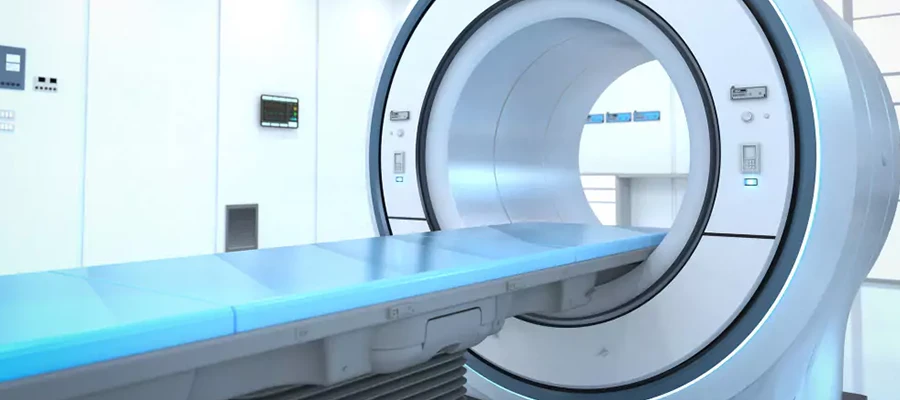
How is a Perfusion MRI Performed?
Patients are placed inside the machine and asked to remain still. Perfusion MRI records signals returning from magnetic fields and radio waves, as well as protons. Contrast images clarify details of vessels and lesions. Non-contrast images may be sufficient in some cases. Metal objects should be removed before the procedure. The images are processed on a computer, and the doctor can perform a detailed analysis. Perfusion MRI plays a critical role in the evaluation of vessels and tissues and is safely used for patients suspected of having tumors, strokes, or vascular occlusions. The scan time generally ranges from 20 to 60 minutes.
Is preparation necessary before a perfusion MRI?
Before a perfusion MRI, all metal objects should be removed and any regular medication should be reported to the doctor. If a contrast-enhanced scan is to be performed, it is recommended to come on an empty stomach. Comfortable clothing will make it easier for the patient to remain still inside the machine. Special conditions such as pregnancy or kidney disease should be reported in advance. Earplugs or headphones can provide comfort due to the noise generated by the machine. Perfusion MRI clarifies vascular and lesion details and plays a critical role in diagnosis and treatment planning. The scan time generally ranges from 20 to 60 minutes and is safely performed in modern medicine.
Is Perfusion MRI Harmful?
Perfusion MRI is safe for most patients because it does not involve radiation, and the contrast agent rarely causes mild side effects. The patient must remain still during the procedure and remove all metal objects. Contrast-enhanced perfusion MRI clarifies details of vessels and lesions, providing accurate diagnosis. Non-contrast scans may be sufficient in some cases. Perfusion MRI is safely applied to critical areas such as the brain, heart, and kidneys, and serves as an important reference in treatment planning. The scan time varies between 20 and 60 minutes, and it is used safely in modern medicine.
Differences Between Perfusion MRI and Diffusion MRI
Perfusion MRI and diffusion MRI are two imaging methods used for different purposes, both examining body tissues in detail. Perfusion MRI provides detailed information about organ function by measuring blood flow and tissue nutrition. Diffusion MRI, on the other hand, shows tissue density and potential damage by analyzing the movement of water molecules within cells. Contrast-enhanced perfusion MRI reveals vascular and lesion details more clearly, facilitating diagnosis in conditions such as tumors or strokes. Non-contrast scans may provide sufficient information in some cases. The patient must remain still during the procedure and remove any metal objects. Perfusion MRI and diffusion MRI results are used reliably in modern medicine to help doctors create treatment plans. The scan time generally ranges from 20 to 60 minutes and provides high-quality image results.
How to Interpret Perfusion MRI Results?
The data obtained provides detailed analyses evaluating blood flow and tissue oxygenation. Contrast-enhanced perfusion MRI clarifies details of vessels and lesions, more clearly indicating suspected tumors, strokes, or vascular occlusions. Non-contrast MRI may be sufficient in some cases. The images are processed on a computer, and the doctor makes an accurate diagnosis by analyzing the blood supply to organs and tissues. The patient must remain still and remove all metal objects during the procedure. Thanks to its detailed data, perfusion MRI provides a reliable reference in diagnosis and treatment planning. This process plays a critical role in modern medicine and contributes to the treatment process of patients.
Things to Consider After Perfusion MRI
Following the procedure, adequate fluid intake helps the contrast agent to be eliminated from the body quickly and supports the healing process. Light exercise or rest may be recommended. Rarely, allergic reactions, skin redness, or itching may occur after contrast-enhanced perfusion MRI. Contact your doctor if you experience any discomfort. Metal objects do not affect the image after the procedure. Perfusion MRI clarifies vascular and lesion details and plays a critical role in diagnosis and treatment planning. Post-procedure precautions contribute to a safe and effective follow-up process. The patient’s position and comfort during the scan improve image quality.
Perfusion MRI Prices 2026
In 2026, perfusion MRI prices typically range between €2000 and €7000.
Does Perfusion MRI Cause Pain?
Most patients do not experience pain during perfusion MRI; only slight discomfort may occur when contrast medium is administered intravenously. Contrast-enhanced scans clarify details of vessels and lesions, and in some cases facilitate diagnosis. Non-contrast MRI may provide sufficient information in some situations. The patient should remain still during the procedure and remove all metal objects. A slight sensation of pressure or cold is considered normal. Perfusion MRI is safely used for accurate diagnosis and treatment planning. The scan usually takes between 20 and 60 minutes and is radiation-free.
How long does a perfusion MRI take?
Perfusion MRI scans typically take between 20 and 60 minutes to complete, although the duration may vary depending on the chosen protocol. Contrast-enhanced scans may take slightly longer to clearly show details of vessels and lesions. Non-contrast MRIs may be completed in a shorter time in some cases. The patient must remain still during the procedure and remove all metal objects. Stillness is critical for image quality and accurate diagnosis. Perfusion MRI is a safe and effective method used in modern medicine and does not involve radiation.
Is Perfusion MRI Safe?
Because it does not involve radiation, perfusion MRI is safe for most patients. The device’s magnetic field operates safely in both contrast and non-contrast scans. The patient must remain still and remove all metal objects during the procedure. Perfusion MRI clarifies vascular and lesion details and provides accurate diagnosis. Mild side effects are rare and usually short-lived. It is safely applied in modern medicine and constitutes a critical reference for accurate diagnosis. The scan takes between 20 and 60 minutes and is completed safely.
What diseases can be diagnosed with perfusion MRI?
Perfusion MRI is used to evaluate diseases such as tumors, strokes, vascular occlusions, and inflammation. Blood flow in critical areas such as the brain, heart, and kidneys is examined in detail. Contrast-enhanced images clarify vessels and lesions, facilitating diagnosis. Non-contrast MRI may be sufficient in some cases. During the procedure, the patient must remain still and remove all metal objects. The images are processed by computer, and the doctor performs an analysis. Perfusion MRI plays a critical role in modern medicine for the early diagnosis and treatment planning of diseases. The scan takes between 20 and 60 minutes.
When will the perfusion MRI results be available?
Perfusion MRI results are usually prepared by a radiologist within 1–3 business days of the scan being completed. In emergency situations, results may be available within a few hours. Contrast-enhanced images show vascular and lesion details more clearly. Non-contrast MRI may be sufficient in some cases. The patient must remain still during the procedure and remove all metal objects. The images are processed on a computer, and the doctor makes a diagnosis by analyzing blood flow and tissue nutrition. Perfusion MRI provides a reliable reference in diagnosis and treatment planning. The scan time varies between 20–60 minutes and it is safely used in modern medicine.