

What is ultrasonography?
Ultrasonography, a crucial method in medical imaging, allows for the examination of the body’s internal structures using sound waves. It works by sending high-frequency sound waves into tissues and converting the reflected signals into images. This method enables detailed examination of organs, soft tissues, and vascular structures. Considered a safe method due to its lack of radiation, ultrasonography is widely used, particularly in pregnancy monitoring. It also plays a significant role in evaluating organs such as the liver, kidneys, gallbladder, and thyroid. Today, advanced equipment allows for clearer and more detailed images, increasing diagnostic accuracy. Due to its rapid application and emphasis on patient comfort, ultrasonography stands out as one of the most preferred imaging methods in healthcare, offering a significant advantage in early diagnosis.
For what purposes is ultrasound used?
Using the correct methods in diagnosis and monitoring processes is of great importance for the early detection of diseases, and ultrasound has a wide range of applications in this regard. Ultrasound is used to examine the structure of internal organs, detect formations such as cysts and tumors, and monitor the pregnancy process. It is also preferred for evaluating vascular structures, examining blood flow, and identifying certain infections. Being a safe method due to its lack of radiation, ultrasound is frequently used, especially in sensitive patient groups. This method is widely preferred in healthcare facilities because it provides quick results and is easy to apply. Thanks to ultrasound, diseases can be detected in the early stages, and the treatment process can be planned more effectively.
How does ultrasound work?
Ultrasonography, one of the fundamental methods in imaging technologies, is based on the principle of the interaction of sound waves with tissues. During ultrasonography, high-frequency sound waves emitted from the device strike body tissues, reflect back, and these reflections are converted into an image by the device. Since different tissues reflect sound waves differently, detailed images are obtained. Thanks to this method, important information about the structure and function of organs is obtained. Ultrasonography also allows for the examination of moving structures thanks to its real-time image provision. Considered a safe method due to its lack of radiation, ultrasonography plays an important role in the diagnostic process and helps in the early detection of many diseases.
In what situations is ultrasound preferred?
Choosing the appropriate imaging method is necessary for accurate disease assessment, and ultrasound is a frequently preferred option. Ultrasound is used particularly for abdominal pain, bloating, pregnancy monitoring, and organ examinations. It also plays an important role in the evaluation of thyroid diseases, kidney problems, and gallstones. Its safety due to not involving radiation is among the reasons for its preference. Ultrasound provides great convenience for both patient and doctor due to its rapid application and immediate results. Therefore, ultrasound is one of the first-choice imaging methods in many different medical conditions.
What diseases can be visualized with ultrasonography?
Early diagnosis is crucial in the treatment of diseases, and ultrasonography stands out as an effective imaging method in this process. Liver diseases, kidney stones, gallbladder problems, and cyst formations can be easily detected with ultrasonography. In addition, thyroid diseases, soft tissue masses, and some vascular diseases can also be examined with ultrasonography. During pregnancy, the development of the baby can also be monitored in detail using this method. Thanks to ultrasonography, diseases can be detected in the early stages, and the treatment process can be started more quickly. Due to its wide range of applications, this method plays an important role in the diagnosis of many different diseases and is frequently preferred in the healthcare field.
What are the types of ultrasound?
With advancements in technology, various types of ultrasound have been developed to meet different needs, and each is used in different fields. These types include abdominal ultrasound, Doppler ultrasound, and transvaginal ultrasound. Abdominal ultrasound is used to examine internal organs, while Doppler ultrasound is preferred for evaluating blood flow. Transvaginal ultrasound is used in the examination of gynecological diseases. Different types of ultrasound offer different advantages depending on their application, and choosing the correct method increases diagnostic accuracy. Thanks to this variety, ultrasound is effectively used in many different medical fields.
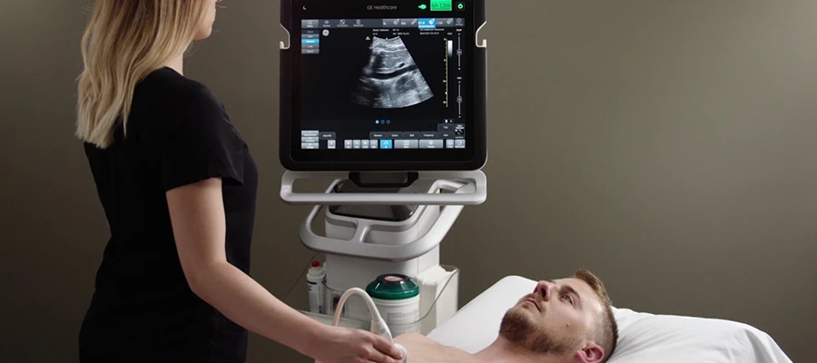
How is an ultrasound scan performed?
For the imaging process to proceed correctly, specific steps must be followed, and the ultrasound scan is performed accordingly. During the ultrasound scan, the patient lies on the examination table, and a special gel is applied to the area to be examined. This gel allows for better transmission of sound waves. Then, a device called a probe is moved across the skin to obtain images. Ultrasound scans are painless and quick. The obtained images are immediately displayed on the screen and evaluated by the doctor. Thanks to its comfortable nature, ultrasound is easily tolerated by patients and plays an important role in the diagnostic process.
Ultrasound Preparation Process
In imaging procedures, preparation may be necessary in some cases to obtain accurate results, and ultrasound has specific guidelines in this regard. Depending on the area being examined, preparations such as fasting or drinking water may be required before the ultrasound. For example, fasting is recommended for abdominal ultrasounds, while a full bladder may be necessary for bladder examinations. Following the doctor’s instructions before the ultrasound improves image quality and ensures more accurate results. This preparation process is a crucial step that directly affects diagnostic accuracy.
How long does an ultrasound take?
Imaging methods that offer a time advantage contribute to the rapid advancement of healthcare services, and ultrasound is quite practical in this respect. An ultrasound procedure is usually completed in 10 to 20 minutes. The total time may be slightly longer with the preparation process, but the procedure itself is quite fast. During the ultrasound, the patient lies comfortably, and the procedure is performed painlessly. The images obtained thanks to ultrasound are evaluated immediately, and the diagnostic process is accelerated. This fast and comfortable process is one of the most important advantages of ultrasound.
Who is a suitable candidate for ultrasound?
Ultrasound, a modern imaging method used to examine the internal structure of the body, is a technique that can be safely applied to a wide range of patients. It is particularly suitable for pregnant women, children, and individuals for whom radiation exposure is contraindicated. Furthermore, ultrasound can be used for detailed examinations in individuals experiencing abdominal pain, bloating, organ dysfunction, and suspected masses. It is also frequently preferred for evaluating organs such as the kidneys, liver, gallbladder, and thyroid. Because it does not involve radiation, it can be safely used for repeated check-ups. Ultrasound increases patient comfort due to its quick and painless application. Therefore, ultrasound is considered one of the first-choice imaging methods in different age groups and for various health problems, making a significant contribution to the diagnostic process.
Things to Consider After an Ultrasound Scan
While no special care is usually required after imaging procedures, paying attention to a few small details can make the process more comfortable, and this is quite simple after an ultrasound. Patients can generally return to their daily lives immediately after an ultrasound because the procedure is non-invasive and does not cause any side effects. Simply rinsing the gel used during the procedure from the skin is sufficient, and no extra care is needed. Rarely, a slight feeling of discomfort may occur after an ultrasound, but this usually resolves on its own. Unless otherwise advised by the doctor, no specific restrictions are applied. Because ultrasound is a safe and fast method, the things to be mindful of after the procedure are quite limited. This provides a significant advantage in terms of patient comfort and allows for a quick return to daily life.
Ultrasound Prices 2026
In 2026, ultrasound prices typically range between €300 and €1500.
Why is an ultrasound performed?
Ultrasound plays a significant role among imaging methods used for the early diagnosis and accurate evaluation of diseases. It is used to examine the structure of internal organs, detect formations such as cysts and tumors, and monitor pregnancy. Detailed examinations can be performed using ultrasound in cases of abdominal pain, bloating, kidney problems, and gallbladder disorders. Ultrasound is also preferred for evaluating vascular structures and blood flow. Being a safe method due to its lack of radiation, ultrasound is frequently used, especially in sensitive patient groups. Thanks to ultrasound, diseases can be identified in the early stages, allowing for more effective treatment planning, which provides a significant advantage in healthcare.
What does an ultrasound show?
Ultrasound holds a significant place among imaging techniques that allow for detailed examination of the body’s internal structure. It clearly shows the structure of organs such as the liver, kidneys, gallbladder, and thyroid. In addition, cysts, tumors, stone formations, and fluid accumulations can be detected with ultrasound. During pregnancy, the development and condition of the baby can also be examined in detail using ultrasound. Furthermore, vascular structures and blood flow can be evaluated with this method. Because ultrasound works on the principle that different tissues reflect sound waves differently, it provides detailed and reliable images. This helps in the early diagnosis of diseases and guides the treatment process.
Is ultrasound safe?
The safety of methods used in healthcare is of great importance to patients, and ultrasound is considered one of the safest methods in this regard. Because it does not involve radiation, ultrasound can be used safely, especially during pregnancy and with children. This procedure, which uses sound waves, does not harm the body and does not cause any side effects. Ultrasound is highly beneficial when applied only when necessary and significantly contributes to the diagnostic process. Therefore, ultrasound is widely used today as a safe and effective imaging method.