

What is scintigraphy?
Scintigraphy, a significant method in nuclear medicine, is an advanced imaging technique used to assess the functional state of organs and tissues in the body. It allows for the examination of organ function through the administration of low doses of radioactive material to the patient. Images obtained through scintigraphy reveal cellular and metabolic activity, aiding in the early diagnosis of diseases. Scintigraphy is frequently used, particularly in the evaluation of organs such as bones, the thyroid, and the heart. It allows for the analysis not only of the disease’s structure but also its functional status. In modern medicine, scintigraphy stands out as a reliable and effective diagnostic method. It offers significant advantages in terms of early diagnosis and accurate treatment planning, playing an active role in the evaluation of many diseases.
For what purposes is scintigraphy used?
Among the methods used in medical diagnostic processes, scintigraphy plays an important role in evaluating organ functions and early diagnosis of diseases. Scintigraphy is particularly used to determine the spread of cancer, evaluate bone diseases, and examine thyroid function. Thanks to scintigraphy, the functioning level of organs is analyzed in detail. Scintigraphy is also an effective method in detecting infections and inflammatory diseases. The data obtained with scintigraphy helps in the correct planning of the treatment process. Scintigraphy is a reliable imaging method with a wide range of uses in modern medicine. By enabling the early detection of diseases, scintigraphy increases the success of treatment.
How does scintigraphy work as a nuclear medicine imaging method?
Thanks to advancements in imaging technologies, scintigraphy stands out as an advanced system for analyzing organ functions. Scintigraphy works by monitoring the distribution of a radioactive substance administered to the patient within the body. During scintigraphy, this substance accumulates in target organs and is imaged by special cameras. The images obtained through scintigraphy provide detailed information about the functioning of the organs. By analyzing metabolic activities, scintigraphy helps in the early diagnosis of diseases. Scintigraphy is widely used in modern medicine as a reliable and effective imaging method.
In what situations is scintigraphy preferred?
Scintigraphy holds a significant place among the methods used for accurate disease assessment. It is particularly preferred in cases of bone pain, thyroid disorders, and suspected cancer. Scintigraphy allows for a clear analysis of disease spread and organ function. It is also used in the evaluation of infections and inflammations. By aiding in accurate diagnosis, scintigraphy facilitates the planning of the treatment process. It offers a significant advantage in early diagnosis and is widely used in modern medicine.
What diseases can be detected with scintigraphy?
Scintigraphy, a diagnostic method, plays an effective role in identifying many different diseases. Bone metastases, thyroid diseases, and heart conditions can be detected with scintigraphy. By analyzing the functional status of organs, the type of disease is clearly revealed. Scintigraphy is a particularly important method for evaluating cancer spread. The results obtained from scintigraphy help guide the treatment process. In modern medicine, scintigraphy stands out as an important diagnostic tool and provides reliable results.
What does bone scintigraphy show?
Among the methods used to assess bone health, scintigraphy offers the possibility of detecting changes in bone tissue at an early stage. Bone scintigraphy reveals conditions such as metastases, fractures, or infections in the bones. Abnormal activities in the bones can be clearly identified thanks to scintigraphy. Bone scintigraphy plays an important role, especially in the assessment of cancer spread. Scintigraphy is an effective method for the early diagnosis of bone diseases.
In what situations is thyroid scintigraphy performed?
Scintigraphy holds a significant place among the methods used to evaluate the functions of the thyroid gland. Thyroid scintigraphy is performed to assess the presence of nodules, hyperthyroidism, and other thyroid diseases. Through scintigraphy, the functioning of the thyroid gland can be analyzed. Thyroid scintigraphy helps in making an accurate diagnosis and in planning the treatment process. Scintigraphy is an effective method in the evaluation of thyroid diseases.
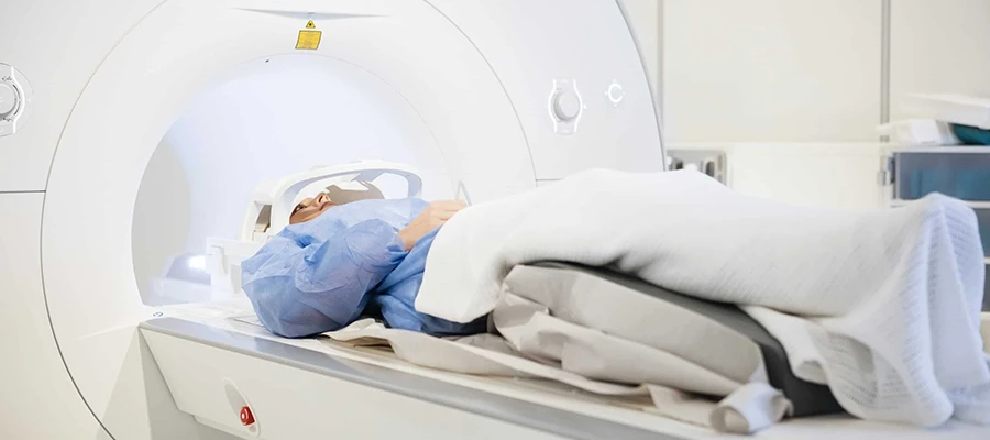
How is a scintigraphy scan performed?
The correct execution of imaging procedures directly affects the quality of the results obtained, and scintigraphy is performed according to specific steps. During scintigraphy, a radioactive substance is administered to the patient, and a certain waiting period is observed. The patient lies still during the scintigraphy scan, and the device captures the images. The scintigraphy procedure is generally completed comfortably and painlessly. Scintigraphy is a controlled and safe imaging method.
Preparation Process Before Scintigraphy
Preparation is crucial for obtaining accurate results from medical procedures, and certain rules must be followed before a scintigraphy scan. The doctor should be informed about any medications being taken before the scan. Fasting may be necessary before the scan in some cases. The preparation process directly affects image quality. With proper preparation, scintigraphy provides more reliable results.
How long does a scintigraphy procedure take?
Thanks to advanced technologies, imaging procedures are completed within a certain time frame, and scintigraphy therefore requires a planned process. A scintigraphy procedure typically takes between 30 minutes and 2 hours. There may be a waiting period for the radioactive substance to distribute throughout the body during the procedure. Since scintigraphy provides a detailed examination, the duration can vary from person to person. Scintigraphy offers a planned process in terms of time.
Is scintigraphy harmful?
Safety is a crucial aspect of medical procedures, and scintigraphy is a method that is applied with great care. Scintigraphy uses a low dose of radiation, but this dose is generally within safe limits. When performed under medical supervision, scintigraphy is considered safe. Given its benefits, scintigraphy has relatively low risks. It is a reliable imaging technique widely used in modern medicine.
Differences Between Scintigraphy and PET CT
Among imaging methods, scintigraphy has different characteristics from other techniques. While scintigraphy evaluates the functions of organs, PET CT offers a more detailed metabolic analysis. Scintigraphy generally focuses on specific organs, while PET CT can perform a whole-body evaluation. Scintigraphy and PET CT are complementary methods used for different purposes. Scintigraphy plays an important role in the evaluation of certain diseases.
How are scintigraphy results interpreted?
One of the crucial stages of the diagnostic process is the accurate evaluation of the data obtained, and scintigraphy results are analyzed by specialist doctors. Scintigraphy results provide detailed information about the functional status of organs. Findings obtained through scintigraphy reveal the presence and spread of disease. Accurate interpretation of scintigraphy results is of great importance for planning the treatment process. With accurate evaluation, scintigraphy is used as an effective diagnostic method.
Scintigraphy Prices 2026
In 2026, scintigraphy prices typically range between €1200 and €4500.
Why is scintigraphy performed?
In medical diagnostic processes, scintigraphy is used to evaluate organ functions and detect diseases in their early stages. Scintigraphy is particularly preferred for diagnosing bone diseases, thyroid problems, and cancer spread. It allows for a detailed analysis of organ function. Scintigraphy is also an effective method for detecting infections and inflammatory diseases. The data obtained through scintigraphy helps in making accurate diagnoses and planning the treatment process. Scintigraphy is a reliable imaging method widely used in modern medicine. It offers a significant advantage in terms of early diagnosis.
What does scintigraphy show?
Scintigraphy, a diagnostic method, provides detailed information about the functional status and metabolic activities of organs. It can detect abnormal changes in bones, the functioning of the thyroid gland, and the spread of certain types of cancer. By analyzing activity at the cellular level, scintigraphy enables the early identification of diseases. The images obtained through scintigraphy provide important information about the spread and severity of the disease. Scintigraphy plays a particularly important role in the detection of metastases. In modern medicine, scintigraphy is a reliable and effective diagnostic method.
Is scintigraphy safe?
Safety is always a top priority in medical procedures, and scintigraphy is a method applied with this in mind. Because the radioactive substances used during scintigraphy are low doses, it is generally considered safe. When performed under medical supervision, scintigraphy has very low risks. Considering the benefits it provides, scintigraphy stands out as a reliable imaging method. Scintigraphy is a safe diagnostic tool widely used in modern medicine.
Does scintigraphy involve radiation?
Scintigraphy, an imaging method, is a technique that involves low levels of radiation. During scintigraphy, images are obtained using a radioactive substance administered to the patient, and this substance is quickly eliminated from the body. In terms of radiation dose, scintigraphy is generally within safe limits. When performed under medical supervision, scintigraphy does not pose a serious health risk. In modern medicine, scintigraphy is considered a safe and effective imaging method.